-

- menu
The urinary system is one of the most important systems in a human organism. Its task is to manage bodily fluids balance by blood filtration and the production of urine, which consists of the filtered waste.
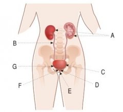 A – kidneys, B – ureters, C – urinary bladder, D – neck of the urinary bladder, E – urethra, F – sphincters, G – nerve endings
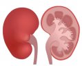 Paired organs which are the most important part of the urinary system. The kidneys produce urine in the process of filtering the blood from toxic substances. Proper functioning of the kidneys is crucial to keep the organism in good condition.
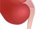 Two thin, smooth muscular tubes; the ureters propel the urine from the kidneys to the urinary bladder. When the bladder is full the ureters close up automatically to prevent the urine from travelling back into the kidneys.
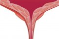
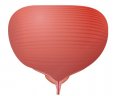 A muscular container placed in the lower abdomen. Its walls are elastic and bundled which allow it to hold from 2 to 3 cups of urine, and with extreme expansion it can hold up to approx 6–8 cups. The bladder wall muscles expand when the bladder is filled, and contract when it is voided.
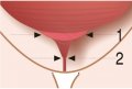
 At the bottom of the bladder there is an internal urethra orifice secured with two sets of ring shaped muscles (sphincters).

 It is the end part of the urinary system. It is a tube with thin walls used to excrete the urine collected in the bladder. The urethra is of different length in men and women due to the differences in their anatomy. The female urethra is about 1–2" cm long, and the male urethra is about 6–8" long and opens at the end of the penis also providing an additional role being the exit for the semen.
 Urine flow is controlled with two sphincters – external and internal. The internal sphincter is a ring shaped muscle which surrounds the initial part of the urethra. It contracts and relaxes involuntarily. The external sphincter is made of the pelvic floor muscles which are spread between the pelvic bones shaped like an inverted umbrella. These muscles are responsible for supporting the organs in the abdominal cavity and are used to control the urine flow voluntary.
 There are receptors in the bladder walls – they react to the tension put on the walls, so they react when the bladder is filled sending a signal to the spinal cord. First urge signals appear after filling the bladder with 1 cup of urine.
Constant kidney functioning regulates electrolytes and maintains acid-base homeostasis. About 170–180 litres of blood flows every day through the kidneys – approx 1,5 litres is excreted in the form of urine. It is a cycle which takes place according the diagram below:
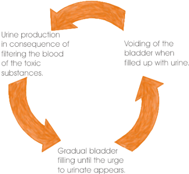
The urine is gathered in the bladder which expands in the process. When there is about 1 cup of urine collected, the receptors in the bladder’s walls send a signal which triggers the urge to urinate. A healthy person with a correctly functioning urinary system is able to control bladder voiding and may postpone this moment if necessary.
Voiding the bladder begins with relaxing of the sphincters and the bladder contraction which pushes the urine outside. The urine flows through the urethra. Then the sphincters contract and the urine begins to fill up the bladder. Urinary system dysfunctions may lead not only to urinary incontinence, but can also have more serious consequences including organism poisoning.
A well-chosen product means both the patient's comfort and less risk of serious pressure sores, as well as less work for the caregiver. Choose a product with our help.
Choose productChoosing the right product is very important.
That is why, we created a simple tool called "Diagnostics".